At OASES, our team of doctors comprise of ophthalmologists that have specialized in ocular oncology thus ensuring all cases are diagnosed and treated in a timely and effective manner.
Patient - 1
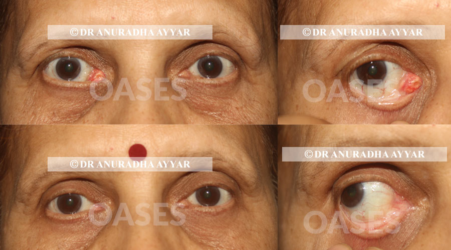
A woman in her fifties was referred for a painless growth in the inner aspect of her right eye since 1 year. Clinically sebaceous gland carcinoma of the caruncle was suspected and the mass was excised completely. The pathology examination confirmed the diagnosis and margins were clear.
The patient was investigated thoroughly considering her past history of two other cancers.
She did well post surgery and is cancer free since 2 years, as of her most recent follow up.
Sebaceous gland carcinoma while is common on the eyelids, is rare in the caruncular region. Early diagnosis and surgery as defined by the protocol for eye cancer, systemic screening and regular follow-up can save lives as well as detect early spread or recurrence of the disease.
Left untreated, this cancer is notorious for systemic spread and death, as also having a potential for recurrence.
Contact UsEye cancer is a type of cancer that impacts the cells of the eye and can occur within the eye, on the surface of the eye, around the orbit, or the eyelids and tear ducts. Based on the area where they occur and the severity of the case, the type and treatment of cancer is diagnosed. At OASES, our team of doctors comprise of ophthalmologists that have specialized in ocular oncology thus ensuring all cases are diagnosed and treated in a timely and effective manner.
This condition refers to a malignancy of the conjunctiva and cornea. It is typically presents as a growth or mass on eye surface and tends to progress slowly. People who are highly exposed to the sun and those who are immunocompromised (transplant patients, with HIV, and other conditions that reduce immunity) are more at risk of developing this condition. Diagnosis requires a physical examination by an ophthalmologist and may require a biopsy for confirmation. In some cases, OSSN can be treated by chemotherapy with Interferons, Mitomycin C eye drops while others may require surgery. The decision is taken after detailed history taking and examination of the eye by the consultant.
In the eyelid region, the first signs of cancer may appear as a mass, thickening or ulceration, or loss of eyelashes. Timely diagnosis and treatment of this cancer is critical to prevent it from spreading to the orbit or elsewhere in the body. The growth may be discolored or even impact the position of the eyelid. In case of any such abnormal appearance of the eyelid, it is essential to consult an Oculoplasty surgeon at the earliest. Diagnosis of this condition can be done on clinical examination and after a biopsy to check whether the growth is benign or malignant. Larger masses will need CT scan/MRI and other investigations as well. Treatment may include surgical removal and eyelid reconstruction. After pathological examination, further treatment in the form of chemotherapy, radiation or even cryotherapy may be discussed.
Cancer that starts inside the eyeball is called intraocular cancer. The most common intraocular cancers are melanoma and retinoblastoma. In general, treatment options for these conditions involve surgery, radiation, and chemotherapy. Retinoblastoma
Retinoblastoma is a cancerous tumor in the retina which is located at the back of the eyeball and is commonly seen in children. The symptoms commonly manifest as inflammation or whitening of the pupil.
Diagnosis requires a physical examination by an ocular oncologist followed by a CT scan and other investigations as may be needed. The treatment will depend on staging and grouping of the cancer. Modalities include surgery, cryotherapy, chemotherapy, radiation. The treatment is a multi-modal and will involve an Ocular oncologist, a paediatric oncologist, paediatrician, radiation oncologist to name a few.
Melanoma is a form of pigmented cells that form a tumor inside the eye. It can be of three types:
Symptoms include pigmentation, poor or blurred vision and a sensation of flashing lights. Diagnosis requires an expert ophthalmologist as a biopsy is not possible in all cases. Based on the size and severity of the case, treatment may include laser therapy, radiation therapy, or surgical excision.
Thus, when it comes to matters of the eye, an examination by a proficient ophthalmologist is important to ensure timely diagnosis and treatment of anything that may be disease causing. Be sure to schedule a routine eye screening every 6 to 12 months to ensure your eyes are healthy and your vision clear. This screening routine is recommended for all age groups from a renowned eye care specialist.
Patient - 2
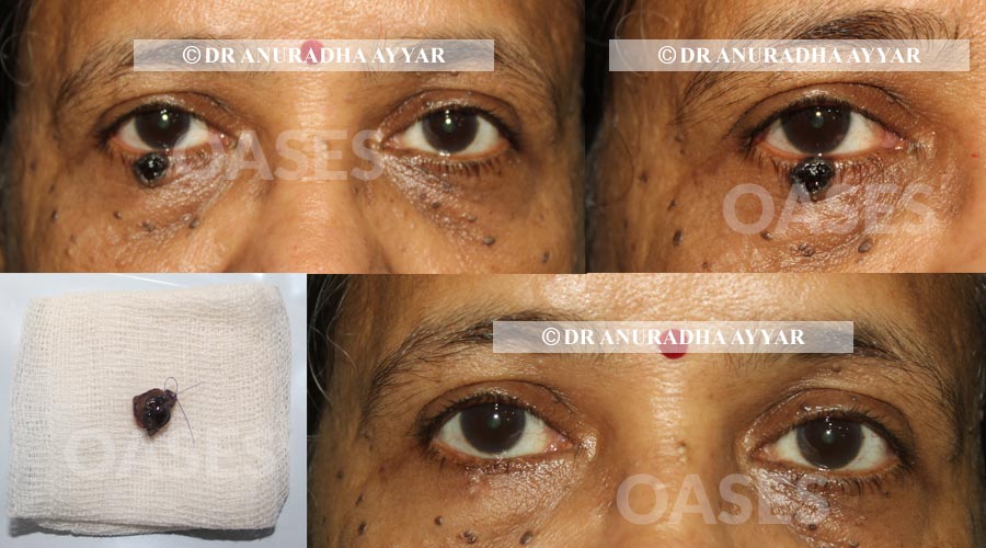
This 49 year old woman was referred for a suspected pigmented basal cellcarcinoma of the right lower eyelid.
We confirmed the diagnosis clinically and planned excision with margins under local anesthesia.
Basal cell carcinoma is a locally invasive cancer and has low rate of systemic spread. Pathology confirmed the diagnosis and declared that the surgical margins were clear of cancer.
Patient did well post surgery with respect to her eyelid on a 2 year follow-up.
She has a history of multiple cancers unfortunately and had to undergo treatment for the same.
Contact UsPatient - 3
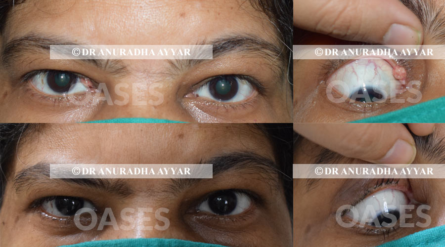
This patient here had been operated for chalazion of the right upper eyelid five times over last 12 years. She had this unusual growth since 6 months now, for which she was referred to Dr Ayyar. We diagnosed her clinically with Right upper eyelid Sebaceous gland carcinoma. She was thoroughly investigated and operated for the eyelid cancer this month. The tissue was examined in detail by the Pathologist and margins were confirmed to be tumor free. Reconstructive surgery for the defect was done with attention to best possible cosmesis whilst maintaining all necessary protocols while dealing with cancerous tissues.
Due to past history of a systemic cancer and family history of multiple cancers, she was evaluated post operatively with a PET SCAN and returned to us with a normal report.
She is officially cancer free now!! And has assured us to come regularly for follow up to detect any early recurrence, if any.
Contact Us